
Hysteroscopic Surgery
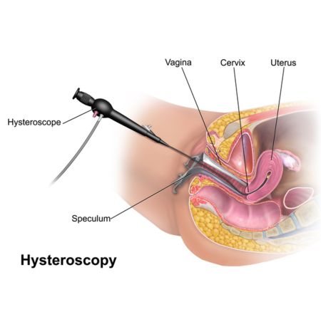
Hysteroscopy is a minimally invasive surgical procedure used to examine the inside of the uterus (the womb). During a hysteroscopy, a thin, lighted tube called a hysteroscope is inserted through the vagina and cervix into the uterus. The hysteroscope has a camera that allows the surgeon to view the uterine cavity on a monitor.
Hysteroscopy can be performed for both diagnostic and therapeutic purposes. For diagnostic hysteroscopy, the surgeon examines the uterine cavity to diagnose conditions such as abnormal bleeding, uterine fibroids, polyps, adhesions (scar tissue), or congenital abnormalities.
In addition to diagnosis, hysteroscopy can also be used for therapeutic procedures. For example, certain conditions identified during the diagnostic hysteroscopy can be treated simultaneously. This may include removal of uterine polyps, fibroids, or adhesions, or performing endometrial ablation (destroying the uterine lining) to treat heavy menstrual bleeding.
TREATMENT
Hysteroscopy is typically performed as an outpatient procedure under local or general anesthesia, depending on the individual’s needs and the complexity of the procedure. Recovery is usually quick, with minimal discomfort, and most women can resume normal activities within a day or two after the procedure. Hysteroscopy is generally considered safe, but as with any surgical procedure, there are risks, including bleeding, infection, or injury to the uterus or surrounding structures.